
Structure Is Not Function: Why OCT Alone Does Not Settle a Glaucoma Question
Imaging shows the anatomy of the optic nerve with great precision. It does not show how much of the signal reaches perception. That gap is why glaucoma care still needs a functional test, and why that test should be easier than it is today.
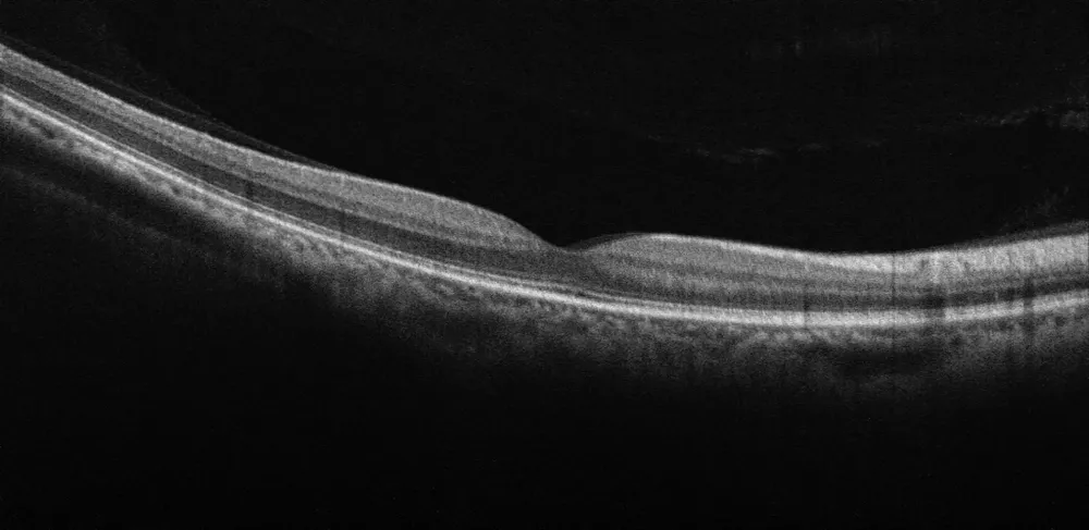
Glaucoma is defined by two things: damage to the optic nerve, and the loss of visual function that follows from it. A diagnosis rests on both. Over the past twenty years the first half has become fast, objective and reproducible, thanks to optical coherence tomography (OCT). The second half is still measured much as it was in the 1980s: a patient in a dark room, a chin rest, and a button to press when a light appears.
This post is about why the second half still matters, and what it would take to make it as routine as the first.
What Imaging Sees
OCT quantifies the retinal nerve fibre layer and the ganglion cell layer to the micrometre. Fundus photography documents the appearance of the optic disc. Both describe the anatomy of the signal source: how many axons are left, how the rim looks, whether the structure has changed since last time.
What they cannot describe is what the patient sees with that structure. The relationship between anatomy and perception is not one to one. Two eyes with a similar nerve fibre layer can have quite different visual fields, and structural and functional measurements often disagree about how far a disease has progressed (Hood and Kardon, 2007). In advanced glaucoma, OCT thickness reaches a floor and stops tracking further loss, while the visual field keeps changing (Mwanza et al., 2015). In early disease the order can go either way: sometimes the structure changes first, sometimes the function (Medeiros et al., 2012).
None of this is a shortcoming of OCT. It is simply that the question “how much does this patient see” is a functional question, and anatomy answers a different one.
Why Function Is Still Measured with a Button
Standard automated perimetry (SAP) remains the reference for visual function in glaucoma, and for good reason: it maps sensitivity location by location across the central field, on a grid that every ophthalmologist can read. But the measurement depends on the patient. They have to hold fixation, keep their attention up for several minutes per eye, decide whether a faint flash was really there, and press a button at the right moment. Reliability indices exist precisely because those conditions are often not met. Results vary between sessions, more so as the damage grows (Artes et al., 2002), and the first test is often treated as practice.
The consequence in daily care is that functional testing happens less often than it should. It is scheduled, it takes a trained perimetrist and a dedicated room, and some patients cannot complete it at all.
Measuring Function Through the Eyes Themselves
SONDA takes a different route to the same question. The patient wears a pair of lightweight glasses with integrated eye-tracking sensors and follows a moving target on a screen. There is no calibration and no button. The result is reliable from a single measurement, completed within two minutes, whatever the severity of the disease.
The measurement is the agreement between where the target is and where the gaze is, moment by moment. Where the target is seen, the eyes follow it. Where it falls on a part of the field that no longer works, the eyes do not react, or react late. Function is read from the oculomotor response, which is involuntary in the sense that matters here: nobody has to decide whether they saw something.
How It Lines Up with the Gold Standard
The cleared use of SONDA is the detection of functional visual field loss as an aid in the diagnosis and staging of primary open-angle glaucoma. The evidence behind it comes from two clinical investigations in glaucoma, summarised on the Clinical evidence page.
In the pivotal investigation (registration NL84477.042.23), the SONDA Score correlated with Zeiss HFA mean sensitivity at r = 0.78 (p < 0.001), across the full severity range. The earlier case-control study (Vrijling et al., 2025a) found the same pattern: tracking performance decreased with glaucoma, most strongly in the severe cases, and the degree of loss on SAP was reflected in the degree of loss in tracking.
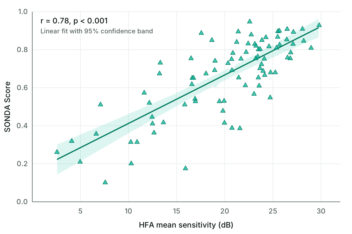
SONDA Score against Zeiss HFA mean sensitivity, pivotal investigation NL84477.042.23. Line: linear fit with its 95% confidence band.
The two instruments rest on entirely different physical principles, one on reported flashes, the other on eye movements, and yet they describe the same loss.
The same relationship can be looked at from the structural side. The figure below plots the SONDA Score against the Disc Damage Likelihood Scale (DDLS) stage of the optic disc, measured by OCT. The median score falls as the structural stage advances, from about 0.75 at stage 4 to about 0.27 at stage 9. The spread within each stage is wide, and adjacent stages overlap considerably: eyes with the same amount of disc damage can differ a great deal in how well they function. That overlap is the structure-function gap in a single picture, and it is why the two measurements complement rather than replace each other.
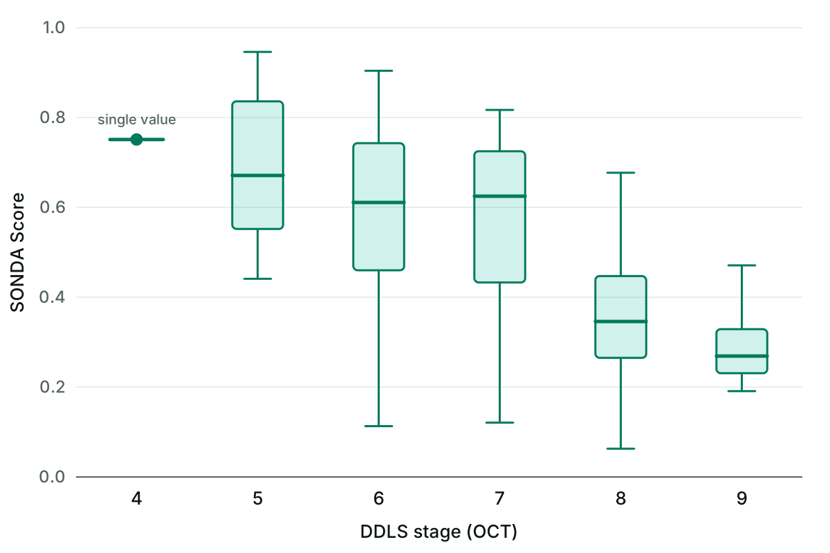
SONDA Score by DDLS stage, pivotal investigation NL84477.042.23. Boxes show the interquartile range, the line the median, whiskers the range. Stage 4 contains a single eye.
Two Measurements of One Disease
Structure and function are two views of the same condition, and neither replaces the other. What has changed is that the functional view no longer has to be the slow one. A functional measurement that is reliable in a single session of under two minutes, needs no dark room and does not depend on the patient’s attention can sit next to the OCT in a routine visit, in the hospital and, as a support for referral decisions, in the optometry practice.
The full validation data, including diagnostic accuracy by stage and test-retest reliability, are on the Clinical evidence page. The published studies are listed under Publications.
References
- Artes, P.H., Iwase, A., Ohno, Y., Kitazawa, Y., Chauhan, B.C. (2002). Properties of perimetric threshold estimates from Full Threshold, SITA Standard, and SITA Fast strategies. Investigative Ophthalmology & Visual Science, 43(8), 2654–2659.
- Hood, D.C., Kardon, R.H. (2007). A framework for comparing structural and functional measures of glaucomatous damage. Progress in Retinal and Eye Research, 26(6), 688–710. doi:10.1016/j.preteyeres.2007.08.001
- Medeiros, F.A., Zangwill, L.M., Bowd, C., Mansouri, K., Weinreb, R.N. (2012). The structure and function relationship in glaucoma: implications for detection of progression and measurement of rates of change. Investigative Ophthalmology & Visual Science, 53(11), 6939–6946. doi:10.1167/iovs.12-10345
- Mwanza, J.C., Budenz, D.L., Warren, J.L., Webel, A.D., Reynolds, C.E., Barbosa, D.T., Lin, S. (2015). Retinal nerve fibre layer thickness floor and corresponding functional loss in glaucoma. British Journal of Ophthalmology, 99(6), 732–737. doi:10.1136/bjophthalmol-2014-305745
- Vrijling, A.C.L., de Boer, M.J., Renken, R.J., Marsman, J.B.C., Heutink, J., Cornelissen, F.W., Jansonius, N.M. (2025a). Detecting and quantifying glaucomatous visual function loss with continuous visual stimulus tracking: a case-control study. Translational Vision Science & Technology, 14(2), 3. doi:10.1167/tvst.14.2.3
- Vrijling, A.C.L., de Boer, M.J., Renken, R.J., Marsman, J.B.C., Heutink, J., Cornelissen, F.W., Jansonius, N.M. (2025b). Screening for glaucoma with a novel eye movement perimetry technique based on continuous visual stimulus tracking. medRxiv preprint, in review. doi:10.1101/2025.10.17.25338104